")

STED

Technique
STED microscopy was awarded with the Nobel prize in chemistry in 2014. It is a superresolution technique that overcomes the diffration limit of light microscopy. In STED microscopy, the diffraction limited excitation spot of a confocal microscope is overlaid with a doughnut-shaped spot of a second laser. The wavelength of the doughnut-shaped laser is choosen in a way that it can de-excite fluorophores which were excited by the standard confocal laser beam. In consequence, only fluorophores in the center of the doughnut (which is much smaller than the diffraction limit) contribute to the fluorescence image. By scanning this "sharpend" laser sopt over the sample a super resoved image can be aquired.
Use
Allows for the acquisiton of super-resolved images. In biological samples, resolution can go down to ~ 25 nm x 25 nm in x,y. Ideal for colocalisation studies due to the fact that both excitation wavelengths are depleted by one STED laser. Bleaching and phototoxicity are the constant companions of superresolution micorscopy, making acquisition of 3D stacks and live cell experiments difficult.
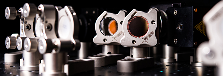
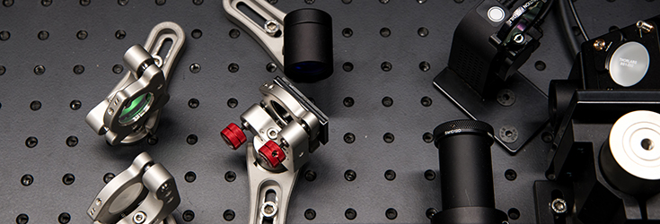
Configuration
Manufacturer:
Abberior Instruments
Type: 3D STED
Microscope body: Olympus
Illumination:
excitation laser
pulsed diode laser 594nm, <500mW (PDL 594, Abberior Instruments)
pulsed diode laser 638nm, 20mW(PiL063X, Advanced Laser Diode Systems)
STED Laser
pulsed fiber laser 775nm, 1200mW (PFL-P-30- 775B1R, MPB Communications)
3D Module
SLM based (easy3D, Abberior Instruments)
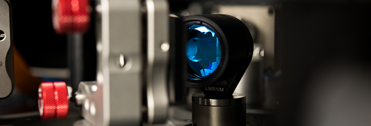
Optics:
100x1.4 Oil UPlanSApo
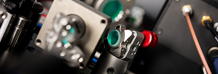
Detectors:
APD: 605nm - 625nm
APD: 650nm - 720nm
Software:
Imspector