")

Spinning disk confocal
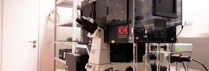
Technique
The difference between a point scanning confocal and a spinning disk confocal microscope is the number of image points acquired at the same time. While a point scanning confocal microscope images one spot at a time, a spinning disk confocal microscope illuminates about 1000 spots in parallel and detects emitted light using a camera. The advatage of this approach is an increase in imaging speed and / or reducion of peak power at the illuminated spot. The latter is important for survival of cells during live cell imaging experiments. Imaging many confocal spots at the same time has the disadvantage of loosing sectioning quality. With increasing density of the confocal spots, the probability of photons emitted from a certain focal spot being detected through the pinhole of a nighboring spot rises.
Use
Confocal technique for investigation of fluorescent living cells. Kinetic studies are possible due to the high speed of this technique. Depending on the configuration of the instrument, microirradiation studies for photoactivation / inactivation are possible.
Configuration
Microscope:
Invers Nikon TiE
Focus hold system:
Ti-ND6-PFS-S Perfect Focus System
Transmittance illumination:
LED 100 coolwhite (can be triggered)
Stage:
Multiwell plate, 35 mm dish and slide format
200 µm z-focus Piezo nano drive
Environmental control:
Large Cage incubator CO2, humidity, heating
Objectives:
CFI P-Achromat 10X/ 0.25/
CFI Apo LWD 40x WI Lambda-S/ 1.15*
CFI P-Apo 40x Lambda/ 0.95*
CFI Apochromat TIRF 60x Öl/ 1.49/*
CFI P-Apo 100x Lambda Öl/ 1.45*
*Differential Interference Contrast (DIC) available
Laser light source:
Andor laser line combiner
Diode 405 nm
DPSS 488 nm
DPSS 561 nm
Diode 638 nm
Spinning disk confocal unit:
Yokogawa W1 spinning disk unit
50 µm disk pattern
Beamsplitter:
Quad band dichroic mirror 405/488/561/640
Pass 400-410, 483-493,557-563,633-643; Reflection 425-470,505-542,578-623, 660-750
Triple band dichroic mirror 445/515/594
Pass 440-450, 513-517,592-596; Reflection 463-497, 532-575, 613-750
Emission Filter:
Multi band
Semrock Brightline, Triple Band Fluorescence Filter, 440-40 / 521-21 / 607-34 / 700-45
Single band
525/50 nm
540/30 nm
600/50 nm
700/75 nm
Polarizer
Borrealis for homogenous illumination
Light stimulation and bleaching (FRAP, FLIP, photactivation)
Frappa with 70:30 quad dichroic 405/488/561/640
Camera:
ANDOR iXon DU-888U3-CS0-BV EMCCD camera
1024 x 1024 pixel
Pixel size 13 x 13 µm
QE >90%
Peltier cooling -85 °C
TIRF:
Nikon TIRF condenser with one motorized axis
Widefield Epifluorescence:
Light source:
LUMENCOR SOLA SE II
Filtercube: (HC BrightLine)
DAPI (ex 390/18; dm 416, em 460/60)
eGFP (ex 469/35, dm 497, em 525/39)
Cy3.5 (ex 565/24, dm 585, em 620/52
Software:
NIS elements
JOBS
Real Time Aquisition
Computer:
Xeon 6Core Prozessor
32 GB RAM
nVidia Quadro K2200 graphic card
512 GB SSD
4 TB SATA Festplatte