")

STED
In the Bioimaging practical course mouse fibroblast cells were used in order to demonstrate the potential of the super-resolution STED technique.
The cells were labeled with antibodies against the nuclear pore protein Nup153 and antibodies against Histone 3 lysine 4 trimethylation (H3K4me3) which is an activation marker for gene expression associated with euchromatin. The cells were fixed with formalin and subsequently imaged with STED microscope.
On figure 1 a comparison between an image done with a Laser Scanning Confocal microscope and a STED microscope of the same cell is shown. The nuclear pores appear in yellow and the euchromatin in blue.
Our results demonstrate a significant difference in resolution between the two images. Further the images prove that in this experiment super-resolution STED microscopy was able to obtain detailed information on cellular structures with a theoretical resolution up to 40 nm and reveal hidden secrets of the cell.
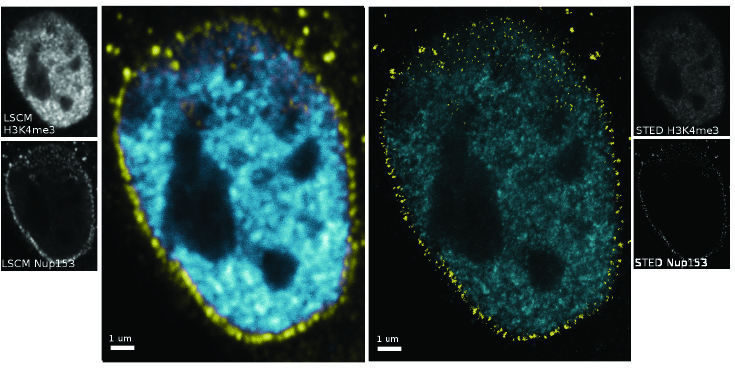
Fig.1: Mouse fibroblast nucleus imaged with Laser Scanning Confocal microscopy and STED.
Confocal (left) and STED (right) images show nuclear pores (yellow) and euchromatin (blue) stained with specific antibodies. Row data pictures (b/w) are shown aside.